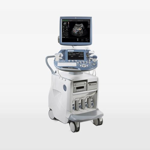
GE Voluson E8, 4-dimensional (4D) detailed ultrasound device is used to follow the development of the baby in the womb during pregnancy. Generally, the device is known as detailed ultrasound or detailed ultrasound. The GE Voluson E8 detailed ultrasound device used in our hospital monitors the development of the baby in the womb by examining its organs such as the brain, eyes, nose, lips, face, neck, heart, lungs, arms, hands, fingers, abdominal organs, back, legs and feet. Thanks to the detailed ultrasonography device that detects the problems related to the formation of these organs, 95 percent of congenital diseases in the womb can be diagnosed in our hospital.
The GE Voluson E8 4-dimensional detailed ultrasound device that we use in our hospital is the most advanced ultrasonography system currently developed in the world. Detailed ultrasound combines 4D ultrasound and black and white ultrasound systems. The most important feature of the 4D system is that it displays images synchronously. The GE Voluson E8 ultrasound device is distinguished from other ultrasound systems with its high-sensitivity endovaginal probe technology. Thanks to this technology, the device provides the opportunity to examine the baby in detail, even in the first trimester. With the help of this technology, anomalies such as spina bifida, cardiac anomalies, chromosomal anomalies, skeletal system disorders can be detected at the beginning of pregnancy. In addition to these, the speckle reduction imaging (SRI) and cross X beam (CRI) features of the device allow images to pass through special filters and allow easier viewing of organs that are difficult to evaluate, such as the heart.
With its spatio-temporal image correlation (STIC) feature, the GE Voluson E8, which can successfully evaluate the heart of the baby in the womb, stands out as an examination method that is becoming more and more common in cardiac evaluation in the world today. Similar to the computerized tomography system, the GE Voluson E8 ultrasound device, which can evaluate images retrospectively, can also make very sensitive measurements in bone structures. In addition, thanks to the GE Voluson E8 4-dimensional detailed ultrasound device, which is considered one of today’s indispensable technologies, the margin of error in in vitro fertilization and infertility treatments and egg measurements is reduced to zero.
Factors such as, ensuring that the birth is carried out under appropriate conditions and planned if there are problems in vital organs, and that the baby is less affected by these problems with detailed ultrasound, intervening in some diseases in the womb and accordingly increasing the chance of survival of the baby, being a guide in diagnosing genetic diseases, thanks to some special ultrasound findings, the position of the baby, determining the position of the baby, the placement of the placenta and the mode of delivery can be shown among the advantages of the GE Voluson E8 ultrasound device.
Detailed ultrasonography can usually be performed between 11th-13th weeks. Detailed ultrasonography performed between these weeks allows the diagnosis of 75% of structural anomalies. However, since it is not possible to detect some problems in brain formation and some holes in the heart during these weeks, it is recommended to repeat the procedure between 20th-24th weeks to evaluate brain development and small holes in the heart.
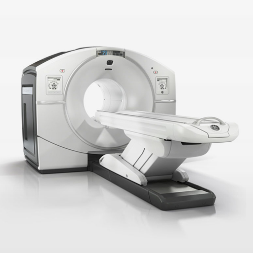
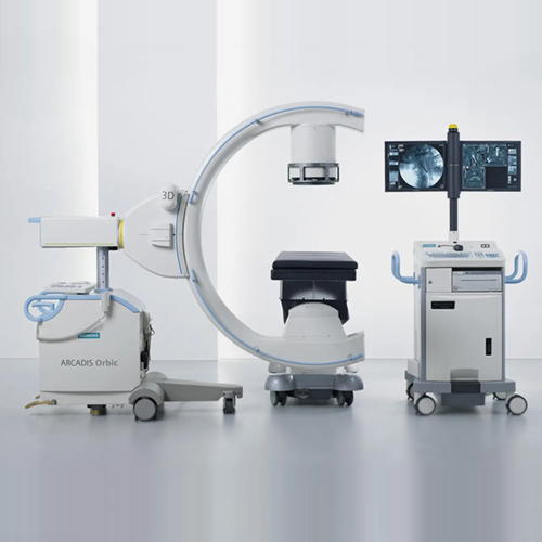
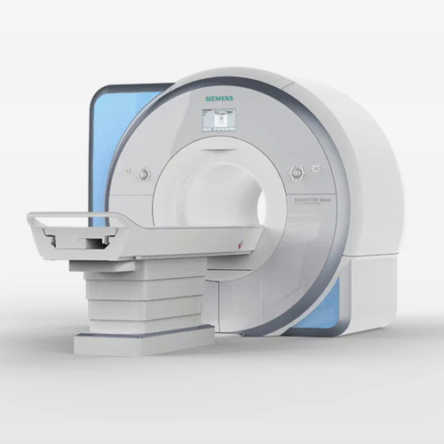
We have all kinds of world-class technical equipment and physical infrastructure that can intervene in our patients, in our hospital. All medical devices in our hospital for diagnosis, treatment, patient care and patient safety are the products of internationally recognized brands.
As Reyap Hospital, we provide uninterrupted and complete service with our large and experienced staff, using up-to-date technological tools in line with up-to-date scientific information.
